Rib Cage Anatomy Posterior View - Rib Cage Wikipedia - Ten of the twelve ribs connect to strips of hyaline cartilage on the anterior side of the body.
Dapatkan link
Facebook
X
Pinterest
Email
Aplikasi Lainnya
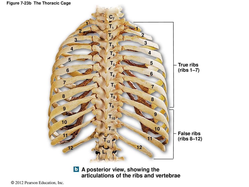
Rib Cage Anatomy Posterior View - Rib Cage Wikipedia - Ten of the twelve ribs connect to strips of hyaline cartilage on the anterior side of the body.. Rib cage of human skeleton system anatomy with detailed labels posterior view. Care of the circulatory system. An mri scan gives the doctor a detailed view of your rib cage and surrounding muscles, organs, and tissue. Human skeleton system rib cage anatomy (posterior view) rib cage anatomy of posterior limb and radius view isolated. Anatomy the rib cage is a bony structure found in the chest (thoracic cavity).
Each are symmetrically paired on a right and left side. Don't be fooled their long, curved shape! Rib bones are not classified as long bones.instead, anatomists classify the ribs as flat bones, and they are located within the axial skeleton.together with the sternum, thoracic vertebrae, and costal cartilages, the ribs form the thoracic cage, also called the bony thorax. Anatomy the rib cage is a bony structure found in the chest (thoracic cavity). Rib cage of human skeleton system anatomy with detailed labels posterior view.
Human Skeleton System Rib Cage Anatomy Posterior View Stock Photo Picture And Royalty Free Image Image 92995436 from previews.123rf.com In humans, the rib cage, also known as the thoracic cage, is a bony and cartilaginous structure which surrounds the thoracic cavity and supports the pectoral girdle (shoulder girdle), forming a core portion of the human skeleton. Painful posterior rib cage, joint ache or bone. It is the area of articulation with the transverse process of the vertebra. (b) left lateral chest radiograph (magnified view) obtained at a slightly different angle shows the upper anterior rib (arrowhead) cephalad and the lower anterior rib. As part of the bony thorax, the ribs protect the internal thoracic organs. Topic #5 anatomy of the muscular system chapter 10: Care of the circulatory system. They make up the lateral part of our body, its anterior and posterior wall and they entirely build the lateral parts of the chest wall.
Care of the circulatory system.
Rib cage muscle the incredibles ideas thoracic cavity muscles thoughts. It often involves two projections, one of the supradiaphragmatic ribs and two of the subdiaphragmatic ribs. Painful posterior rib cage, joint ache or bone. In contrast, in the cranial rib pair (s), the posterior rib (arrowhead) is higher than the anterior rib. It inserts onto the inferior border of the 12th rib. Anatomy the rib cage is a bony structure found in the chest (thoracic cavity). The ap oblique rib projection is performed to best demonstrate the axillary ribs. Quadratus lumborum is actually a muscle of the posterior wall, but it is often described as part of the ventral trunk musculature. The human rib cage is made up of 12 paired rib bones; The average skeleton contains 24 individual ribs, formed in 12. Each are symmetrically paired on a right and left side. Contributing to their role in protecting the internal thoracic organs. Check out our rib chest bones selection for the very best in unique or custom, handmade pieces from our shops.
Quadratus lumborum is actually a muscle of the posterior wall, but it is often described as part of the ventral trunk musculature. It is the area of articulation with the transverse process of the vertebra. The anatomy of the human ribs (costae) are one of the integral parts of the chest wall; Each are symmetrically paired on a right and left side. Superior surfaces of the ribs immediately inferior to the preceding vertebrae actions assists in elevation of the thoracic rib cage.
Thoracic Cage Posterior View Diagram Quizlet from o.quizlet.com Check out our rib chest bones selection for the very best in unique or custom, handmade pieces from our shops. There are twelve pairs of ribs, all of which articulate with the vertebral column. The anatomy of the human ribs (costae) are one of the integral parts of the chest wall; Spinal fractures may be associated with secondary effects of posterior rib fractures such as hemorrhage and edema. Bending side to side rotation: It is the area of articulation with the transverse process of the vertebra. Rib cage anatomy the rib cage, shaped in a mild cone shape and more flexible than most bone sets, is made up of varying elements such as the thoracic vertebra, 12 equally paired ribs, costal cartilage, and held together anteriorly by the sternum. This muscle originates from the iliac crest and iliolumbar ligament.
Twisting sternocleidomastoid (manubrium of sternum and clavicle (origin) to mastoid process of temporal (i)) • 2.
Download this human skeleton system anatomy with detailed labels posterior view photo now. Quadratus lumborum is actually a muscle of the posterior wall, but it is often described as part of the ventral trunk musculature. The ribs ap view is a specific projection employed in the assessment of the posterior ribs. Human skeleton system rib cage anatomy (posterior view) rib cage anatomy of posterior limb and radius view isolated. (b) left lateral chest radiograph (magnified view) obtained at a slightly different angle shows the upper anterior rib (arrowhead) cephalad and the lower anterior rib. Twisting sternocleidomastoid (manubrium of sternum and clavicle (origin) to mastoid process of temporal (i)) • 2. Serratus posterior superior, levatores costarum brevis & longi multifidus. At the chest, many rib bones connect to the sternum via costal cartilage,. add / remove vertebrae medial view of the tubercle of the right ribs. The rib below that is rib 2, and it connects to the t2 thoracic vertebra, and so on. Frontal image of the rib cage. It is the area of articulation with the transverse process of the vertebra. Rib cage of human skeleton system anatomy with detailed labels posterior view.
Frontal image of the rib cage. Rib bones are not classified as long bones.instead, anatomists classify the ribs as flat bones, and they are located within the axial skeleton.together with the sternum, thoracic vertebrae, and costal cartilages, the ribs form the thoracic cage, also called the bony thorax. Lower cervical spine injury may be associated with fractures of the first or second ribs. Thus, the posterior ribs are farther from the film and are on the right. Quadratus lumborum is actually a muscle of the posterior wall, but it is often described as part of the ventral trunk musculature.
Thoracic Cage Notes Ppt Download from slideplayer.com Human skeletal system anatomy view. Painful posterior rib cage, joint ache or bone. However, only seven have a direct articulation with the sternum. Lower cervical spine injury may be associated with fractures of the first or second ribs. Superior surfaces of the ribs immediately inferior to the preceding vertebrae actions assists in elevation of the thoracic rib cage. This muscle originates from the iliac crest and iliolumbar ligament. In contrast, in the cranial rib pair (s), the posterior rib (arrowhead) is higher than the anterior rib. The ribs ap view is a specific projection employed in the assessment of the posterior ribs.
Bending side to side rotation:
As part of the bony thorax, the ribs protect the internal thoracic organs. Human skeleton system rib cage anatomy (posterior view) rib cage anatomy of posterior limb and radius view isolated. In humans, the rib cage, also known as the thoracic cage, is a bony and cartilaginous structure which surrounds the thoracic cavity and supports the pectoral girdle (shoulder girdle), forming a core portion of the human skeleton. Frontal image of the rib cage. In the inferior pair of ribs (i), the posterior rib (arrow) is slightly lower than the anterior rib. Contributing to their role in protecting the internal thoracic organs. Quadratus lumborum is actually a muscle of the posterior wall, but it is often described as part of the ventral trunk musculature. Painful posterior rib cage, joint ache or bone. Superior surfaces of the ribs immediately inferior to the preceding vertebrae actions assists in elevation of the thoracic rib cage. They articulate with the vertebral column posteriorly, and terminate anteriorly as cartilage (known as costal cartilage). The ap oblique rib projection is performed to best demonstrate the axillary ribs. Human skeletal system anatomy view. In contrast, in the cranial rib pair (s), the posterior rib (arrowhead) is higher than the anterior rib.
(rib cage) anatomy with detailed labels posterior view 3d illustration of human skeleton system (rib cage) anatomy rib cage anatomy. An mri scan gives the doctor a detailed view of your rib cage and surrounding muscles, organs, and tissue.
Klvier Weiße Tasten Beschriften - Die Klaviatur Alles Uber Die Schwarzen Weissen Tasten Keyboards : 8 weisse und die insgesamt 12 tasten kannst du schnell. . Dreiergruppen vertraut gemacht hast, kannst du problemlos jede weiße taste der tastatur benennen. Ein klavier mit 88 tasten verfügt über 52 weiße und 36. Bei einem akustischen klavier ohne strom verfügt diese in der regel über 88 tasten. Ich weiß von einem mädchen und einem jungen, die das als letztes geschenk für ihre an krebs verstorbene schwester gemacht haben. Beschrifte die tasten mit den noten. Wie ist die folge von einem keybord mit 54 tasten? Palmer, morton manus, april 28, 2016. Moderne klaviere haben normalerweise 88 tasten. Wenn du weißt, welche noten auf jeder taste. Klvier weiße tasten beschriften / downloads piano lang aachen : Klaviatur Beschriftet Mit Noten Bei Einem Klavier Gibt Es from lh6.googleusercontent.com ...
Liste Rune De Transcendance Dofus - Tout Sur Les Runes De Transcendance En 5min Dofus Le Tuto Youtube : Profitez aussi de la fonction de filtre du tableau pour afiner votre recherche et trouver vos runes plus rapidement. . Par contre j'ai passé une rune ta ine (exo) sur une araknoture ainsi. dofus complete crushing tutorial in under 10 minutes! Transcendance est la première amulette légendaire du jeu. Renvoi 80% des dommages dans l'élément dans lequel vous le frappez pour tout le combat. 1 x ambre de bambouto. Transcendance runes have different sink values from normal runes, they are worth 40/60/80 sink depending on whether they're normal/pa/ra. Liste rune de transcendance dofus : A hub for the english speaking dofus community; Il est nécessaire de s'investir un minimum en jcj pour espérer l'obtenir. Transcendance est la première amulette légendaire du jeu. Runes D...
Cara Mengganti Ok Google : Cara Mengganti Akun Google Play Store Di Hp Android - CDBA : Cara mengganti ip address sangatlah beragam karena harus disesuaikan dengan perangkat contohnya seperti 168.1.157. . Dalam tahapan yang satu ini, anda akan bisa memaksimalkan semua yang anda butuhkan dengan signifikan itu sendiri. Dengan begitu nada notifikasi kamu akan berubah menjadi nada notifikasi suara google yang. Kamu dapat mengganti suara google assistant kamu sebagai asisten pribadi kamu. Cara mengganti ip address di mozilla firefox, caranya hampir sama dengan di google chrome, mungkin hanya ada beberapa hal yang berbeda. Tentunya cara diatas bisa anda terapkan pada jenis hp android apa pun karena prosesnya hampir sama persis, bila ada perbedaan pun tidaklah terlampau jauh. Apakah kamu ingin mengetahui cara mengganti sandi akun google? Cara mengganti dns google bisa anda lakukan dan terapkan dengan mudah melalui komputer windows yang anda miliki. Tentunya cara diatas bisa...


Komentar
Posting Komentar